Resolución de estructuras cristalográficas
DOI:
https://doi.org/10.3989/arbor.2015.772n2004Palabras clave:
problema de la fase, constricciones, factores de estructura, mapas de Fourier, métodos de búsqueda, minimización, máxima verosimilitud, optimización, restricciones, rayos-XResumen
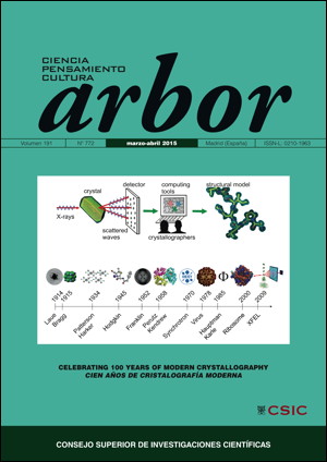
La cristalografía proporciona una visión tridimensional de las moléculas a un nivel de detalle atómico, que no sólo resulta muy informativa sino que además puede ser fácil e intuitivamente comprendida por seres tan predominantemente visuales como solemos ser los humanos. Sin embargo, al contrario que la microscopía, esta técnica no ofrece directamente una imagen y el modelo estructural no puede calcularse directamente a partir de los datos de difracción, ya que solamente las intensidades de los rayos difractados y no sus fases son accesibles a la medida experimental. Para determinar la estructura tridimensional las fases deben ser obtenidas por medio de métodos adicionales, bien experimentales o computacionales. Esto constituye el problema de la fase en cristalografía. En este artículo ofreceremos una visión general de los principales hitos en la búsqueda de las fases perdidas.
Descargas
Citas
Abad-Zapatero, C., Abdel-Meguid, S. S., Johnson, J. E., Leslie, A. G., Rayment, I., Rossmann, M. G., Suck, D. and Tsukihara, T. (1980). Structure of southern bean mosaic virus at 2.8Å resolution. Nature, 286, pp. 33–39. http://dx.doi.org/10.1038/286033a0
Adams, M. J., Blundell, T. L., Dodson, E. J., Dodson, G. G., Vijayan, M., Baker, E. N., Harding, M. M., Hodgkin, D. C., Rimmer, B. and Sheat, S. (1969). Structure of Rhombohedral 2 Zinc Insulin Crystals. Nature, 224, pp. 491. http://dx.doi.org/10.1038/224491a0
Allen, F. H. (2002). The Cambridge Structural Database: a quarter of a million crystal structures and rising. Acta Crystallographica, B58, pp. 380-388. http://dx.doi.org/10.1107/S0108768102003890
Amunts, A., Brown, A., Bai, X. C., Llácer, J. L., Hussain, T., Emsley, P., Long, F., Murshudov, G., Scheres, S. H. and Ramakrishnan V. (2014). Structure of the yeast mitochondrial large ribosomal subunit. Science, 343, pp. 1485-1489. http://dx.doi.org/10.1126/science.1249410 PMid:24675956 PMCid:PMC4046073
Armstrong, H. E. (1927). Poor Common Salt! Nature, 120, pp. 478.
Astbury, W. T. and Street, A. (1932). A. X-Ray studies of the structure of hair, wool, and related fibers. I. General. Philosophical Transactions of the Royal Society A, 230, pp. 75– 101.
Berman, H. M., Westbrook, J., Feng, Z., Gilliland, G., Bhat, T. N., Weissig, H., Shindyalov, I. N. and Bourne, P. E. (2000). The Protein Data Bank. Nucleic Acids Research, 28, pp. 235-242. http://dx.doi.org/10.1093/nar/28.1.235 PMid:10592235 PMCid:PMC102472
Bernal, J. D. and Crowfoot, D. (1934). X-Ray Photographs of Crystalline Pepsin. Nature, 133, pp. 794–795. http://dx.doi.org/10.1038/133794b0
Bernstein, F. C., Koetzle, T. F., Williams, G. J. B., Meyer, E. F., Brice, M. D., Rodgers, J. R., Kennard, O., Shimanouchi, T. and Tasumi, M. (1977). The Protein Data Bank: a computer-based archival file for macromolecular structures. Journal of Molecular Biology, 112, pp. 535-542. http://dx.doi.org/10.1016/S0022-2836(77)80200-3
Bibby, J., Keegan, R. M., Mayans, O., Winn, M. D. and Rigden, D. J. (2012). AMPLE: a cluster-and-truncate approach to solve the crystal structures of small proteins using rapidly computed ab initio models. Acta Crystallographica, D68, pp. 1622-1631. http://dx.doi.org/10.1107/S0907444912039194 PMid:23151627
Bragg, W. L. (1913a). The Diffraction of Short Electromagnetic Waves by a Crystal. Mathematical Proceedings of the Cambridge Philosophical Society, 17, pp. 43–57.
Bragg, W. L. (1913b). The Structure of Some Crystals as indicated by their Diffraction of X-rays. Proceedings of the Royal Society of London, 89, pp. 248-277. http://dx.doi.org/10.1098/rspa.1913.0083 http://dx.doi.org/10.1098/rspa.1913.0083
Bragg, W. L. (1925). The Crystalline Structure of Inorganic Salts. Nature, 116, pp. 557. http://dx.doi.org/10.1038/116249a0
Bragg, W. H. and Bragg, W. L. (1913a). The Reflexion of X-rays by Crystals. Proceedings of the Royal Society of London A, 88, 605, pp. 428–438.
Bragg, W. H. and Bragg, W. L. (1913b). The structure of the diamond. Nature, 91, pp. 557.
Broenniman, C., Eikenberry, E. F., Henrich, B., Horisberger, G., Huelsen, G., Pohl, E., Schmitt, B., Schulze-Briese, C., Suzuki, M., Tomizaki, T., Toyokawa, A. and Wagner, A. (2006) The Pilatus 1M detector. Journal of Synchrotron Radiation, 13, pp. 120-133. http://dx.doi.org/10.1107/S0909049505038665 PMid:16495612
Bunkóczi, G., McCoy, A. J., Echols, N., Grosse-Kunstleve, R. W., Adams, P. D., Holton, J. M., Read, R. J. and Terwilliger, T. C. (2014). Macromolecular X-ray structure determination using weak, single-wavelength anomalous data. Nature Methods, 12, pp. 127–130. http://dx.doi.org/10.1038/nmeth.3212 PMid:25532136 PMCid:PMC4312553
Burla, M. C., Carrozzini, B., Cascarano, G. L., Giacovazzo, C., Polidori, G. (2012). VLD algorithm and hybrid Fourier syntheses. Journal of Applied Crystallography, 45, pp. 1287-1294. http://dx.doi.org/10.1107/S0021889812041155
Caliandro, R., Carrozzini, B., Cascarano, G. L., De Caro, L., Giacovazzo, C. and Siliqi, D. (2005a). Phasing at resolution higher than the experimental resolution. Acta Crystallographica, D61, 556-565.
Caliandro, R., Carrozzini, B., Cascarano, G. L., De Caro, L., Giacovazzo, C., Siliqi, D. (2005b). Ab initio phasing at resolution higher than experimental resolution. Acta Crystallographica, D61, pp. 1080-1087.
Caliandro, R., Carrozzini, B., Cascarano, G. L., De Caro, L., Giacovazzo, C., Mazzone, A., Siliqi, D. (2008). Crystal structure solution of small-to-medium-sized molecules at non-atomic resolution. Journal of Applied Crystallography, 41, pp. 548-553. http://dx.doi.org/10.1107/S002188980800945X
Champness, J. N., Bloomer, A. C., Bricogne, G., Butler, P. G. and Klug, A. (1976). The structure of the protein disk of tobacco mosaic virus to 5A resolution. Nature, 259, pp. 20-24. http://dx.doi.org/10.1038/259020a0 PMid:1250335
Cowtan, K. D. and Main, P. (1993). Improvement of macromolecular electron- density maps by the simultaneous application of real and reciprocal space constraints. Acta Crystallographica, D49, pp. 148-157.. http://dx.doi.org/10.1107/S0907444992007698 PMid:15299555
Crowfoot, D., Bunn, C. W., Rogers-Low, B. W. and Turner-Jones, A. (1949). X-ray crystallographic investigation of the structure of penicillin. In Clarke, H. T., Johnson, J. R. and Robinson, R. (eds). Chemistry of Penicillin. Princeton University Press, pp. 310–367.
Crowther, R. A. and Blow, D. W. (1967) A Method of Positioning a Known Molecule in an Unknown Crystal Structure. Acta Crystallographica, 23, pp. 544-548. http://dx.doi.org/10.1107/S0365110X67003172
Dauter Z. and Adamiak D. A. (2001). Anomalous signal of phosphorus used for phasing DNA oligomer: importance of data redundancy. Acta Crystallographica, D57, pp. 990–995. http://dx.doi.org/10.1107/S0907444901006382
Dauter, Z., Dauter, M., de La Fortelle, E., Bricogne, G. and Sheldrick, G. M. (1999). Can anomalous signal of sulfur become a tool for solving protein crystal structures? Journal of Molecular Biology, 289, pp. 83–92.
DeTitta, G. T., Weeks, C. M. , Thuman, P., Miller, R. and Hauptman, H. A. (1994). Structure solution by minimal function phase refinement and Fourier filtering: theoretical basis. Acta Crystallographica, A50, pp. 203-210. http://dx.doi.org/10.1107/S0108767393008980
DiMaio, F., Terwilliger, T. C., Read, R. J., Wlodawer, A., Oberdorfer, G., Wagner, U., Valkov, E., Alon, A., Fass, D., Axelrod, H. L., Das, D., Vorobiev, S. M., Iwai, H., Pokkuluri, P. R. and Baker, D. (2011). Improved molecular replacement by density- and energy-guided protein structure optimization. Nature, 473, pp. 540-543. http://dx.doi.org/10.1038/nature09964 PMid:21532589 PMCid:PMC3365536
Ewald, P. P. (1913). About the theory of the interference of X-rays in crystals (Zur Theorie der interferenzen der Röntgen-strahlen in kristallen). Physikalische Zeitschrift, 14, pp. 465–472.
Franklin, R. and Gosling, R. G. (1953). Molecular Configuration in Sodium Thymonucleate. Nature, 171, pp. 740-741. http://dx.doi.org/10.1038/171740a0 PMid:13054694
Friedrich, W., Knipping, P. and Laue, M. (1912). Interferenz-Erscheinungen bei Röntgenstrahlen. Sitzungsberichte der Königlich Bayerische Akademie der Wissenschaften, pp. 303–322.
Fujinaga, M. and Read, R. J. (1987). Experiences with a new translation-function program. Journal of Applied Crystallography, 20, pp. 517-521. http://dx.doi.org/10.1107/S0021889887086102
Garman, E. F. and Schneider, T. R. (1997). Macromolecular Cryocrystallography. Journal of Applied Crystallography, 30, pp. 211-237. http://dx.doi.org/10.1107/S0021889897002677
Giacovazzo, C. (2011). Fundamentals of crystallography (3rd ed). Oxford, New York: Oxford University Press - International Union of Crystallography texts on crystallography. http://dx.doi.org/10.1093/acprof:oso/9780199573653.001.0001
Glykos, N. M. and Kokkinidis, M. (2003). Structure determination of a small protein through a 23-dimensional molecular-replacement search. Acta Crystallographica, D59, pp. 709-718. http://dx.doi.org/10.1107/S0907444903002889
Green, D. W., Ingram, V. M. and Perutz, M. F. (1954). Structure of haemoglobin: IV. Sign determination by the isomorphous replacement method. Proceedings of the Royal Society of London. Series A, 225, pp. 287-307. http://dx.doi.org/10.1098/rspa.1954.0203
Hauptman, H. and Karle, J. (1953). Solution of the phase problem I. The centrosymmetric crystal. Dayton, Ohio: American Crystallographic Association.
Harker, D. (1936). The application of the three-dimensional Patterson method and the crystal structures of proustite, Ag3AsS3, and pyrargyrite, Ag3SbS3. The Journal of Chemical Physics, 4, pp. 381-390. http://dx.doi.org/10.1063/1.1749863
Harker, D. (1956). The determination of the phases of the structure factors of non-centrosymmetric crystals by the method of double isomorphous replacement. Acta Crystallographica, 9, pp. 1-9.. http://dx.doi.org/10.1107/S0365110X56000012
Harker, D. and Kasper, J. S. (1948). Phases of Fourier coefficients directly from crystal diffraction data. Acta Crystallographica, 1, pp. 70-75. http://dx.doi.org/10.1107/S0365110X4800020X
Harrison, S. C., Olson, A. J., Schutt, C. E., Winkler, F. K. and Bricogne, G. (1978). Tomato bushy stunt virus at 2.9 Å resolution. Nature, 276, pp. 368-373.
Hendrickson, W. A. (1991). Determination of Macromolecular Structures from Anomalous Diffraction of Synchrotron Radiation. Science, 254, pp. 51-58. http://dx.doi.org/10.1126/science.1925561 PMid:1925561
Hendrickson, W. A. and Teeter, M. M. (1981). Structure of the hydrophobic protein crambin determined directly from the anomalous scattering of sulphur. Nature, 290, pp. 107-113. http://dx.doi.org/10.1038/290107a0
Hodgkin, D. C., Kamper, J., Mackay, M., Pickworth, J., Trueblood, K. N. and White, J. G. (1956). Structure of vitamin B-12. Nature, 178, pp. 64–66. http://dx.doi.org/10.1038/178064a0 PMid:13348621
Hope, H. (1990). Crystallography of biological macromolecules at ultra-low temperature. Annual Review of Biophysics and Biophysical Chemistry, 19, pp. 107-126. http://dx.doi.org/10.1146/annurev.bb.19.060190.000543 PMid:2194473
Huber, R. (1965). Die automatisierte Faltmoleku.lmethode. Acta Crystallographica, 19, pp. 353-356. http://dx.doi.org/10.1107/S0365110X65003444
Jia-Xing, Y., Woolfson, M. M., Wilson, K. S. and Dodson, E. J. (2005). A modified ACORN to solve protein structures at resolutions of 1.7 Å or better. Acta Crystallographica, D61, pp. 1465-1475.
Karle, J. and Hauptman, H. (1950). The phases and magnitudes of the structure factors. Acta Crystallographica, 3, pp. 181-187. http://dx.doi.org/10.1107/S0365110X50000446
Karle, J. and Hauptman, H. (1956). A theory of phase determination for the four types of non-centrosymmetric space groups 1P222, 2P22, 3P12, 3P22. Acta Crystallographica, 9, pp. 635-651. http://dx.doi.org/10.1107/S0365110X56001741
Kendrew, J. C., Bodo, G., Dintzis, H. M., Parrish, R. G., Wyckoff, H. and Phillips, D. C. (1958). A three dimensional model of the myoglobin molecule obtained by X-ray analysis. Nature, 181, pp. 662-666. http://dx.doi.org/10.1038/181662a0 PMid:13517261
Kissinger, C. L., Gehlhaar, D. K. and Fogel, D. B. (1999). Rapid automated molecular replacement by evolutionary search. Acta Crystallographica, D55, pp. 484-491. http://dx.doi.org/10.1107/S0907444998012517
von Laue, M. (1912). Eine quantitative pru.fung der theorie fu.r die interferenz-erscheinungen bei Röntgenstrahlen. Sitzungsberichte der Königlich Bayerische Akademie der Wissenschaften, pp. 363-373.
Liu, Q., Dahmane, T., Zhang, Z., Assur, Z., Brasch, J., Shapiro, L., Mancia, F. and Hendrickson, W. A. (2012). Structures from Anomalous Diffraction of Native Biological Macromolecules. Science, 336, pp. 1033-1037. http://dx.doi.org/10.1126/science.1218753 PMid:22628655 PMCid:PMC3769101
Loll, P. J., Bevivino A. E., Korty B. D. and Axelsen P. H. (1997). Simultaneous Recognition of a Carboxylate-containing Ligand and an Intramolecular Surrogate Ligand in the Crystal Structure of an Asymmetric Vancomycin Dimer. Journal of the American Chemical Society, 119, pp. 1516-1522.
McCoy A. J., Grosse-Kunstleve R. W., Storoni L. C. and Read R. J. (2005). Likelihood- enhanced fast translation functions. Acta Crystallographica, D61, pp. 458-464. http://dx.doi.org/10.1107/S0907444905001617 PMid:15805601
McCoy A. J., Grosse-Kunstleve R. W., Adams P. D., Winn M. D., Storoni L. C. and Read R. J. (2007). Phaser crystallographic software. Journal of Applied Crystallography, 40, pp. 658-674. http://dx.doi.org/10.1107/S0021889807021206 PMid:19461840 PMCid:PMC2483472
Miller, R., DeTitta, G. T., Jones, R. , Langs, D. A., Weeks, C. M. and Hauptman, H. A. (1993). On the application of the minimal principle to solve unknown structures. Science, 259, pp. 1430-1433. 8451639 http://dx.doi.org/10.1126/science.8451639 PMid:8451639
Mueller, M., Wang, M. and Schulze-Briese, C. (2012). Optimal fine £X-slicing for single-photon-counting pixel detectors. Acta Crystallographica, D68, pp. 42–56. http://dx.doi.org/10.1107/S0907444911049833 PMid:22194332 PMCid:PMC3245722
Oszlanyi, G. and Su.to, A. (2004). Ab initio structure solution by charge flipping. Acta Crystallographica, A60, pp. 134-141. http://dx.doi.org/10.1107/S0108767303027569 PMid:14966324
Patterson, A. L. (1935). A direct method for the determination of the components of interatomic distances in crystals. Zeitschrift fu.r Kristallographie, 90, pp. 517-542. http://dx.doi.org/10.1524/zkri.1935.90.1.517
Pauling, L. and Corey, R. B. (1951). The pleated sheet, a new layer configuration of polypeptide chains. Proceedings of the National Academy of Sciences of the USA, 37, pp. 251-256. http://dx.doi.org/10.1073/pnas.37.5.251 PMid:14834147 PMCid:PMC1063350
Pauling, L., Corey, R. B. and Branson, H. R. (1951). The structure of proteins, two hydrogen-bonded helical configurations of the polypeptide chain. Proceedings of the National Academy of Sciences of the USA, 37, pp. 205-211. http://dx.doi.org/10.1073/pnas.37.4.205 http://dx.doi.org/10.1073/pnas.37.4.205
Proepper, K., Meindl, K., Sammito, M., Dittrich, B., Sheldrick, G. M., Pohl, E. and Uson, I. (2014). Structure solution of DNA-binding proteins and complexes with ARCIMBOLDO libraries. Acta Crystallographica, D70, pp. 1743-1757. http://dx.doi.org/10.1107/S1399004714007603 PMid:24914984 PMCid:PMC4051508
Qian, B., Raman, S., Das, R., Bradley, P., McCoy, A. J., Read, R. J. and Baker, D. (2007). High-resolution structure prediction and the crystallographic phase problem. Nature, 450, pp. 259-64. http://dx.doi.org/10.1038/nature06249 PMid:17934447 PMCid:PMC2504711
Robertson, M. P. and Scott, W. G. (2008). A general method for phasing novel complex RNA crystal structures without heavy-atom derivatives. Acta Crystallographica, D64, pp. 738-744. http://dx.doi.org/10.1107/S0907444908011578 PMid:18566509 PMCid:PMC2507861
Rodríguez, D., Grosse, C., Himmel, S., González, C., Martínez de Ilarduya, I., Becker, S., Sheldrick, G. M. and Usón, I. (2009). Crystallographic ab initio protein solution below atomic resolution. Nature Methods, 6, pp. 651-653. http://dx.doi.org/10.1038/nmeth.1365 PMid:19684596
Rossmann, M. G. (1972). The Molecular Replacement Method. New York: Gordon & Breach.
Rossmann, M. G. and Blow, D. M. (1962) The Detection of Subunits within the Crystallographic Asymmetric Unit. Acta Crystallographica, 15, pp. 24-31. http://dx.doi.org/10.1107/S0365110X62000067
Sammito, M. D., Millán, C., Rodríguez, D., M. de Ilarduya, I., Meindl, K., De Marino, I., Petrillo, G., Buey, R. M., de Pereda, J. M., Zeth, K., Sheldrick, G. M. and Usón, I. (2013). Exploiting tertiary structure through local folds for crystallographic phasing. Nature Methods, 10, pp. 1099-1101. http://dx.doi.org/10.1038/nmeth.2644 PMid:24037245
Sayre, D. (1952). The squaring method: a new method for phase determination. Acta Crystallographica, 5, pp. 60-65. http://dx.doi.org/10.1107/S0365110X52000137
Schäfer, M., Schneider, T. R. and Sheldrick, G. M. (1996). Crystal structure of vancomycin. Structure, 4, pp. 1509-1515. http://dx.doi.org/10.1016/S0969-2126(96)00156-6
Sheldrick, G. M.and Gould, R. (1995). Structure solution by iterative peaklist optimization and tangent expansion in space group P1. Acta Crystallographica, B51, pp. 423-431. http://dx.doi.org/10.1107/S0108768195003661
Sheldrick, G. M. (2008). A short history of SHEL. Acta Crystallographica, A64, pp. 112-122. http://dx.doi.org/10.1107/S0108767307043930 PMid:18156677
Sheldrick, G. M. (2010). Experimental phasing with SHELXC/D/E: combining chain tracing with density modification. Acta Crystallographica, D66, pp. 479-485. http://dx.doi.org/10.1107/S0907444909038360 PMid:20383001 PMCid:PMC2852312
Sheldrick, G. M., Gilmore, C. J., Hauptman, H. A., Weeks, C. M., Miller, R. and Usón, I. (2011). Ab initio phasing. In Arnold, E., Himmel, D. M. and Rossmann, M. G. (eds.). International Tables for Crystallography. Dordrecht: Kluwer Academic Publishers, pp. 413-429.
Storoni L. C., McCoy A. J. and Read R. J. (2004). Likelihood-enhanced fast rotation functions. Acta Crystallographica, D60, pp. 432-438. http://dx.doi.org/10.1107/S0907444903028956 PMid:14993666
Wang, B.-C. (1985). Resolution of phase ambiguity in macromolecular crystallography. Methods in Enzymology, 115, pp. 90-112. http://dx.doi.org/10.1016/0076-6879(85)15009-3
Wang, A. H.-J., Quigley, G. J., Kolpak, F. J., Crawford, J. L., van Boom, J. H., van der Marel, G. and Rich, A. (1979). Molecular structure of a left-handed double helical DNA fragment at atomic resolution. Nature, 282, pp. 680-686. http://dx.doi.org/10.1038/282680a0 PMid:514347
Watson, J. D. and Crick, F. H. C. (1953). Molecular structure of nucleic acids: A structure for deoxyribose nucleic acid. Nature, 171, pp. 737-738. http://dx.doi.org/10.1038/171737a0 PMid:13054692
Weeks, C. M., Adams, P. D., Berendzen, J., Bru.nger, A. T., Dodson E. J., Grosse-Kunstleve, R. W., Schneider, T. R., Sheldrick, G. M., Terwilliger, T. C., Turkenburg, M. and Usón, I. (2003). Automatic Solution of Heavy-Atom Substructures. Methods in Enzymology, 374, pp. 37-83. http://dx.doi.org/10.1016/S0076-6879(03)74003-8
Weinert, T., Olieric, V., Waltersperger, S., Panepucci, E., Chen, L., Zhang, H., Zhou, D., Rose, J., Ebihara, A., Kuramitsu, S., Li, D., Howe, N., Schnapp, G., Pautsch, A., Bargsten, K., Prota, A. E., Surana, P., Kottur, J., Nair, D. T., Basilico, F., Cecatiello, V., Pasqualato, S., Boland, A., Weichenrieder, O., Wang, B.-C., Steinmetz, M. O., Caffrey, M and Wang, M. (2015). Fast native-SAD phasing for routine macromolecular structure determination. Nature Methods, 12, pp. 131-133. http://dx.doi.org/10.1038/nmeth.3211 PMid:25506719
Yang, C., Pflugrath, J. W., Courville, D. A., Stence, C. N. and Ferrara, J. D. (2003). Away from the edge: SAD phasing from the sulfur anomalous signal measured in-house with chromium radiation. Acta Crystallographica, D59, pp. 1943–1957. http://dx.doi.org/10.1107/S0907444903018547
Publicado
Cómo citar
Número
Sección
Licencia
Derechos de autor 2015 Consejo Superior de Investigaciones Científicas (CSIC)

Esta obra está bajo una licencia internacional Creative Commons Atribución 4.0.
© CSIC. Los originales publicados en las ediciones impresa y electrónica de esta Revista son propiedad del Consejo Superior de Investigaciones Científicas, siendo necesario citar la procedencia en cualquier reproducción parcial o total.Salvo indicación contraria, todos los contenidos de la edición electrónica se distribuyen bajo una licencia de uso y distribución “Creative Commons Reconocimiento 4.0 Internacional ” (CC BY 4.0). Puede consultar desde aquí la versión informativa y el texto legal de la licencia. Esta circunstancia ha de hacerse constar expresamente de esta forma cuando sea necesario.
No se autoriza el depósito en repositorios, páginas web personales o similares de cualquier otra versión distinta a la publicada por el editor.


