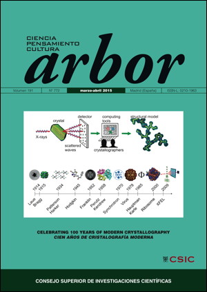
Cristalización de proteínas en el diseño de fármacos en los últimos 50 años
DOI:
https://doi.org/10.3989/arbor.2015.772n2008Palabras clave:
diseño de fármacos, cristalizaciónResumen
Vivimos en una época en la que esperamos ir al médico y obtener una pastilla para curar cualquier dolencia que padezcamos; por desgracia, esta expectativa no es real. Aunque muchos de los remedios en uso provienen de fuentes naturales, la mayoría de los nuevos medicamentos son el resultado de la investigación científica. En el proceso de diseño y descubrimiento de fármacos, la cristalografía de proteínas juega un papel central. Los conocimientos que han hecho esto posible han venido evolucionando desde hace cincuenta años aproximadamente. Los métodos de cristalización de macromoléculas y la determinación de sus estructuras a través de la cristalografía de rayos X han sido automatizados y miniaturizados y la velocidad de la adquisición de datos de difracción ha aumentado en varios órdenes de magnitud. Si hace cincuenta años la resolución de una sola estructura podría llevar varios años, actualmente se pueden determinar las estructuras de varios complejos proteína-ligando en un solo día. La cristalografía de alto rendimiento hoy día es un gran recurso en el proceso del descubrimiento de fármacos pues proporciona una manera rápida y precisa de adaptar los fármacos candidatos a las dianas mediante el análisis de su modo de unión. La cristalización sigue siendo el principal desafío.
Descargas
Citas
Antoni, C., Vera, L., Devel, L., Catalani, M. P., Czarny, B., Cassar-Lajeunesse, E., Nuti, E., Rossello, A., Dive, V. and Stura, E. A. (2013). Crystallization of bi-functional ligand protein complexes. Journal of Structural Biology, 182, pp. 246–254. http://dx.doi.org/10.1016/j.jsb.2013.03.015 PMid:23567804
Berman, H., Henrick, K., Nakamura, H. and Markley, J. L. (2007). The worldwide Protein Data Bank (wwPDB): ensuring a single, uniform archive of PDB data. Nucleic Acids Research, 35, pp. D301–D303. http://dx.doi.org/10.1093/nar/gkl971 PMid:17142228 PMCid:PMC1669775
Chayen, N. E., Shaw Stewart, P. D. and Baldock, P. (1994). New developments of the IMPAX small-volume automated crystallization system. Acta Crystallographica, D50, pp. 456–458. http://dx.doi.org/10.1107/S0907444993013320 PMid:15299401
Chayen, N. E. (1999). Crystallization with oils: a new dimension in macromolecular crystal growth. Journal of Crystal Growth, 196, pp. 434–441. http://dx.doi.org/10.1016/S0022-0248(98)00837-9
Ciccone, L., Tepshi, L., Nencetti, S. and Stura, E. A. (2015). Transthyretin complexes with curcumin and bromo-estradiol: evaluation of solubilizing multicomponent mixtures. New Biotechnology, 32, pp. 54-64. http://dx.doi.org/10.1016/j.nbt.2014.09.002 PMid:25224922
Crowfood, D. and Riley, D. (1939). X-Ray Measurements on Wet Insulin Crystals. Nature, 144, pp. 1011–1012. http://dx.doi.org/10.1038/1441011a0
Cudney, R., Patel, S., and McPherson, A. (1994). Crystallization of macromolecules in silica gels. Acta Crystallographica, D50, pp. 479–483. http://dx.doi.org/10.1107/S090744499400274X PMid:15299406
Dale, G. E., Oefner, C. and D'Arcy, A. (2003). The protein as a variable in protein crystallization. Journal of Structural Biology, 142, pp. 88–97. http://dx.doi.org/10.1016/S1047-8477(03)00041-8
Das, K., Sarafianos, S. G., Clark, A. D., Boyer, P. L. Hughes, S. H. and Arnold, E. (2007). Crystal structures of clinically relevant Lys103Asn/Tyr181Cys double mutant HIV-1 reverse transcriptase in complexes with ATP and non-nucleoside inhibitor HBY 097. Journal of Molecular Biology, 365, pp. 77–89. http://dx.doi.org/10.1016/j.jmb.2006.08.097 PMid:17056061
Giegé, R. (2013). A historical perspective on protein crystallization from 1840 to the present day. FEBS Journal, 280, pp. 6456–97. http://dx.doi.org/10.1111/febs.12580 PMid:24165393
Haupt, H. and Heide, K. (1966). Crystallization of prealbumin from human serum. Experientia, 22, pp. 449–451. http://dx.doi.org/10.1007/BF01900976 PMid:4960491
Howard, J. A. K. (2003). Dorothy Hodgkin and her contributions to biochemistry. Nature Reviews Molecular Cell Biology, 4, pp. 891-896. http://dx.doi.org/10.1038/nrm1243 PMid:14625538
Ivanova, M. I., Sievers, S., Sawaya, M. R., Wall, J. S. and Eisenberg, D. (2009). Molecular basis for insulin fibril assembly. Proceedings of National Academy of Sciences of the USA, 106, pp. 18990–18995. http://dx.doi.org/10.1073/pnas.0910080106 PMid:19864624 PMCid:PMC2776439
Kaldor, S. W., Kalish, V. J., Davies, J. F. 2nd., Shetty, B. V., Fritz, J. E., Appelt, K., Burgess, J. A.,, Campanale, K. M., Chirgadze, N. Y., Clawson, D. K., Dressman, B. A., Hatch, S. D., Khalil, D. A., Kosa, M. B., Lubbehusen, P. P., Muesing, M. A., Patick, A. K., Reich, S. H., Su, K. S. and Tatlock, J. H. (1997). Viracept (nelfinavir mesylate, AG1343): a potent, orally bioavailable inhibitor of HIV-1 protease. Journal of Medicinal Chemistry, 21, pp. 3979-3985. http://dx.doi.org/10.1021/jm9704098 PMid:9397180
Laurent, T. C. (1963). The interaction between polysaccharides and other macromolecules. 5. The Solubility of Proteins in the presence of dextran. Biochemical Journal, 89, pp. 253–257.
Luecke, H., Schobert, B., Richter, H. T., Cartailler, J. P. and Lanyi, J. K. (1999). Structure of bacteriorhodopsin at 1.55 Å resolution. Journal of Molecular Biology, 291, pp. 899-911. http://dx.doi.org/10.1006/jmbi.1999.3027 PMid:10452895
Li, D., Howe, N., Dukkipati, A., Shah, S. T. A., Bax, B. D., Edge, C., Bridges, A., Hardwicke, P., Singh, O. M. P., Giblin, G., Pautsch, A., Pfau, R., Schnapp, G., Wang, M., Olieric, V. and Caffrey, M. (2014). Crystallizing Membrane Proteins in the Lipidic Mesophase. Experience with Human Prostaglandin E2 Synthase 1 and an Evolving Strategy. Crystal Growth & Design, 14, pp. 2034–2047.
Livnah, O., Stura, E. A., Johnson, D. L., Middleton, S. A., Mulcahy, L. S., Wrighton, N. C., Dowr, W. J., Jolliffe, L. K. and Wilson, I. A. (1996). Functional mimicry of a protein hormone by a peptide agonist: the EPO receptor complex at 2.8 Å. Science, 273, pp. 464–71. http://dx.doi.org/10.1126/science.273.5274.464
Naylor, H. M. and Newcomer, M. E. (1999). The structure of human retinol-binding protein (RBP) with its carrier protein transthyretin reveals an interaction with the carboxy terminus of RBP. Biochemistry, 38, pp. 2647-2653. http://dx.doi.org/10.1021/bi982291i PMid:10052934
Nencetti, S. and Orlandini, E. (2012). TTR fibril formation inhibitors: is there a SAR? Current Medicinal Chemistry, 19, pp. 2356–2379.
Otálora, F., Gavira, J. A., Ng, J. D., García-Ruiz, J. M. (2009). Counterdiffusion methods applied to protein crystallization. Progress in Biophysics and Molecular Biology, 101, pp. 26–37. http://dx.doi.org/10.1016/j.pbiomolbio.2009.12.004 PMid:20018206
Palmer, I. and Wingfield, P. T. (2004). Preparation and extraction of insoluble (inclusion- body) proteins from Escherichia coli. Current Protocols in Protein Science, Chapter 6, Unit 6.3. http://dx.doi.org/10.1002/0471140864.ps0603s38 PMid:18429271 PMCid:PMC3518028
Purdy, R. H., Woeber, K. H., Holloway, M. T. and Ingbar, S. H. (1965). Preparation of Crystalline Thyroxine-binding Prealbumin from Human Plasma. Biochemistry, 4, pp. 1888–1895. http://dx.doi.org/10.1021/bi00885a029
Quintas, A., Saraiva, M. J. and Brito, R. M. (1997). The amyloidogenic potential of transthyretin variants correlates with their tendency to aggregate in solution. FEBS Letters, 418, pp. 297–300. http://dx.doi.org/10.1016/S0014-5793(97)01398-7
Stura, E. A. and Wilson, I. A. (1990). Analytical and production seeding techniques. Methods, 1, pp. 38–49. http://dx.doi.org/10.1016/S10462023(05)801458
Stura, E. A. and Wilson, I. A. (1991). Applications of the streak seeding technique in protein crystallization. Journal of Crystal Growth, 110, pp. 270–282. http://dx.doi.org/10.1016/0022-0248(91)90896-D
Stura, E. A., Charbonnier, J. and Taussig, M. J. (1999). Epitaxial jumps. Journal of Crystal Growth, 196, pp. 250–260. http://dx.doi.org/10.1016/S0022-0248(98)00832-X
Syed, R. S., Reid, S. W., Li, C., Cheetham, J. C., Aoki, K. H., Liu, B., Zhan, H., Osslund, T. D., Chirino, A. J., Zhang, J., Finer-Moore, J., Elliot, S., Sitney, K., Katz, B. A., Matthews, D. J., Wendoloski, J. J., Egrie, J. and Stroud, R. M. (1998). Efficiency of signalling through cytokine receptors depends critically on receptor orientation. Nature, 395, pp. 511–516. http://dx.doi.org/10.1038/26773 PMid:9774108
Teeter, M. M. and Hendrickson, W. A. (1979). Highly ordered crystals of the plant seed protein crambin. Journal of Molecular Biology, 127, pp. 219–223. http://dx.doi.org/10.1016/0022-2836(79)90242-0
Thiessen, K. J. (1994). The use of two novel methods to grow protein crystals by microdialysis and vapor diffusion in an agarose gel. Acta Crystallographica, D50, pp. 491–495. http://dx.doi.org/10.1107/S0907444994001332 PMid:15299408
Vera, L., Antoni, C., Devel, L., Czarny, B., Cassar-Lajeunesse, E., Rossello, A., Dive, V. and Stura, E. (2013). Screening Using Polymorphs for the Crystallization of Protein–Ligand Complexes. Crystal Growth & Design, 13, pp. 1878-1888. http://dx.doi.org/10.1021/cg301537n
Ward, K. B., Wishner, B. C., Lattman, E. E. and Love, W. E. (1975). Structure of deoxyhemoglobin a crystals grown from polyethylene glycol solutions. Journal of Molecular Biology, 98, pp. 161–177. http://dx.doi.org/10.1016/S0022-2836(75)80107-0
Whittingham, J. L., Jonassen, I., Havelund, S., Roberts S. M., Dodson E. J., Verma C. S., Wilkinson A. J. and Dodson G. G. (2004). Crystallographic and solution studies of N-lithocholyl insulin: a new generation of prolonged-acting human insulins. Biochemistry, 25, pp. 5987-5995. http://dx.doi.org/10.1021/bi036163s PMid:15147182
Publicado
Cómo citar
Número
Sección
Licencia
Derechos de autor 2015 Consejo Superior de Investigaciones Científicas (CSIC)

Esta obra está bajo una licencia internacional Creative Commons Atribución 4.0.
© CSIC. Los originales publicados en las ediciones impresa y electrónica de esta Revista son propiedad del Consejo Superior de Investigaciones Científicas, siendo necesario citar la procedencia en cualquier reproducción parcial o total.Salvo indicación contraria, todos los contenidos de la edición electrónica se distribuyen bajo una licencia de uso y distribución “Creative Commons Reconocimiento 4.0 Internacional ” (CC BY 4.0). Puede consultar desde aquí la versión informativa y el texto legal de la licencia. Esta circunstancia ha de hacerse constar expresamente de esta forma cuando sea necesario.
No se autoriza el depósito en repositorios, páginas web personales o similares de cualquier otra versión distinta a la publicada por el editor.


