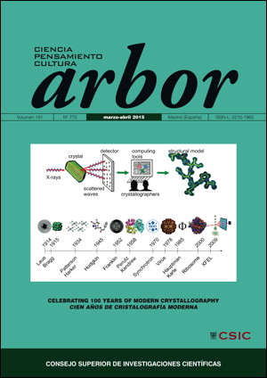
From crystallography to structural biology, a century of discoveries
DOI:
https://doi.org/10.3989/arbor.2015.772n2003Keywords:
biophysics, crystallization, electron microscopy, macromolecular complexes, molecular biology, structural biology, X-ray crystallographyAbstract
From crystallography, the technique mostly used to study the structure of matter, the field mutated into structural biology, has mutated in life sciences into structural biology, which has been developed as an essential and rather successful area of research to fully understand the workings of cellular pathways. The application of physical approaches to biological systems has been crucial to comprehend the structure and function of the biological components of living organisms. In this assay the author walks the reader through the last century, which has witnessed how this life sciences research area was born and moved towards larger assemblies in the core of crucial biological problems. The influence of research in physics, biochemistry and molecular biology has been key in the successes and large body of seminal results obtained by structural biologists. The author proposes that the future of this area implies the integration of its results at the cellular level apart of using more quantitative approaches to describe biological processes.
Downloads
References
Ben-Shem, A., Garreau de Loubresse, N., Melnikov, S., Jenner, L., Yusupova, G., Yusupov, M. (2011). The structure of the eukaryotic ribosome at 3.0 Å resolution. Science, 334, pp. 1524-1529.
Berger, I., Fitzgerald, D. J. and Richmond, T. J. (2004). Baculovirus expression system for heterologous multiprotein complexes. Nature Biotechnology, 22, pp. 1583-1587.http://dx.doi.org/10.1038/nbt1036 PMid:15568020
Bernal, J. D. and Crowfoot, D. (1934). X-Ray photographs of crystalline pepsin. Nature, 133, pp. 794-795.http://dx.doi.org/10.1038/133794b0
Blake, C. C. F., Koenig, D. F., Mair, G. A., North, A. C. T., Phillips, D. C. and Sarma, V. R. (1965). Structure of Hen Egg-White Lysozyme: A Three-dimensional Fourier Synthesis at 2 Å Resolution. Nature, 206, pp. 757-761.http://dx.doi.org/10.1038/206757a0 PMid:5891407
Chance, R. E., Kroeff, E. P., Hoffmann, J. A. and Frank, B. H. (1981). Chemical, physical, and biologic properties of biosynthetic human insulin. Diabetes Care, 4, pp. 147–154.http://dx.doi.org/10.2337/diacare.4.2.147 PMid:7011716
Chapman, H. N., Fromme, P., Barty, A., White, T. H., Kirian, R. A., Aquila, A., Hunter, M. S., Schulz, J., DePonte, D. P., Weierstall, U., Doak, R. B., Maia, F. R. N. C. Martin, A. V., Schlichting, I., Lomb, L., Coppola, N., Shoeman, R. L., Epp, S. W., Hartmann, R., Rolles, D., Rudenko, A., Foucar, L., Kimmel, N., Weidenspointner, G., Holl, P., Liang, M., Barthelmess, M., Caleman, C., Boutet, S., Bogan, M. J., Krzywinski, J., Bostedt, C., Bajt, S., Gumprecht, L., Rudek, B., Erk, B., Schmidt, C., Hömke, A., Reich, C., Pietschner, D., Strüder, L., Hauser, G., Gorke, H., Ullrich, J., Herrmann, S., Schaller, G., Schopper, F., Soltau, H., Kühnel, K. U., Messerschmidt, M., Bozek, J. D., Hau-Riege, S. P., Frank, M., Hampton, C. Y., Sierra, R. G., Starodub, D., Williams, G. J., Hajdu, J., Timneanu, N., Seibert, M. M., Andreasson, J., Rocker, A., Jönsson, O., Svenda, M., Stern, S., Nass, K., Andritschke, R., Schröter, C. D., Krasniqi, F., Bott, M., Schmidt, K. E., Wang, X., Grotjohann, I., Holton, J. M., Barends, T. R. M., Neutze, R., Marchesini, S., Fromme, R., Schorb, S., Rupp, D., Adolph, M., Gorkhover, T., Andersson, I., Hirsemann, H., Potdevin, G., Graafsma, H., Nilsson, B., Spence, J. C. H. (2011). Femtosecond X-ray protein nanocrystallography. Nature, 470, pp. 73-77.http://dx.doi.org/10.1038/nature09750 PMid:21293373 PMCid:PMC3429598
Clemons, W. M. Jr., Brodersen, D. E., McCutcheon, J. P., May, J. L., Carter, A. P., Morgan-Warren, R. J., Wimberly, B. T. and Ramakrishnan, V. (2001). Crystal structure of the 30 S ribosomal subunit from Thermus thermophilus: purification, crystallization and structure determination. Journal of Molecular Biology, 310, pp. 827-843.http://dx.doi.org/10.1006/jmbi.2001.4778 PMid:11453691
Cramer, P., Bushnell, D. A., Fu, J., Gnatt, A. L., Maier-Davis, B., Thompson, N. E., Burgess, R. R., Edwards, A. M., David, P. R. and Kornberg, R. D. (2000). Architecture of RNA polymerase II and implications for the transcription mechanism. Science, 288, pp. 640-649.http://dx.doi.org/10.1126/science.288.5466.640 PMid:10784442
Edwards, A. M., Darst, S. A., Feaver, W. J., Thompson, N. E., Burgess, R. R. and Kornberg, R. D. (1990). Purification and lipid-layer crystallization of yeast RNA polymerase II. Proceedings of the National Academy of Sciences of the USA, 87, pp. 2122-2126.http://dx.doi.org/10.1073/pnas.87.6.2122 PMid:2179949 PMCid:PMC53638
Franklin, R. E. and Gosling, R. G. (1953). Molecular Configuration of Sodium Thymonucleate. Nature, 171, pp. 740-741. http://dx.doi.org/10.1038/171740a0http://dx.doi.org/10.1038/171740a0
Goeddel, D. V., Kleid, D. G., Bolivar, F., Heyneker, H. L., Yansura, D. G., Crea, R., Hirose, T., Kraszewski, A., Itakura, K. and Riggs, A. D. (1979). Expression in Escherichia coli of chemically synthesized genes for human insulin. Proceedings of the National Academy of Sciences of the USA, 76, pp. 106-110.http://dx.doi.org/10.1073/pnas.76.1.106 PMid:85300 PMCid:PMC382885
Grutter, M. G., Marki, W. and Walliser, H. P. (1985). Crystals of the complex between recombinant N-acetyleglin c and subtilisin. Journal of Biological Chemistry, 260, pp. 11436–11437.
Itakura, K., Hirose, T., Crea, R., Riggs, A. D., Heyneker, H. L., Bolivar, F. and Boyer, H. W. (1977). Expression in Escherichia coli of a chemically synthesized gene for the hormone somatostatin. Science, 198, pp. 1056-1063.http://dx.doi.org/10.1126/science.412251 PMid:412251
Johansson, L. C., Arnlund, D., White, T. A., Katona, G., DePonte, D. P., Weierstall, U., Doak, R. B., Shoeman, R. L., Lomb, L., Malmerberg, E., Davidsson, J., Nass, K., Liang, M., Andreasson, J., Aquila, A., Bajt, S., Barthelmess, M., Barty, A., Bogan, M. J., Bostedt, C., Bozek, J. D., Caleman, C., Coffee, R., Coppola, N., Ekeberg, T., Epp, S. W., Erk, B., Fleckenstein, H., Foucar, L., Graafsma, H., Gumprecht, L., Hajdu, J., Hampton, C. Y., Hartmann, R., Hartmann, A., Hauser, G., Hirsemann, H., Holl, P., Hunter, M. S., Kassemeyer, S., Kimmel, N., Kirian, R. A., Maia, F. R. N. C., Marchesini, S., Martin, A. V., Reich, C., Rolles, D., Rudek, B., Rudenko, A., Schlichting, I., Schulz, J., Seibert, M. M., Sierra, R. G., Soltau, H., Starodub, D., Stellato, F., Stern, S., Stru.der, L., Timneanu, N., Ullrich, J., Wahlgren, W. Y., Wang, X., Weidenspointner, G., Wunderer, C., Fromme, P., Chapman, H. N., Spence, J. C. H. and Neutze, R. (2012). Lipidic phase membrane protein serial femtosecond crystallography. Nature Methods, 9, pp. 263-265.http://dx.doi.org/10.1038/nmeth.1867 PMid:22286383 PMCid:PMC3438231
Jordan, P., Fromme, P., Witt, H. T., Klukas, O., Saenger, W. and Krauss, N. (2001). Three-dimensional structure of cyanobacterial photosystem I at 2.5 Å resolution. Nature, 411, pp. 909-917.http://dx.doi.org/10.1038/35082000 PMid:11418848
Kendrew, J. C., Bodo, G., Dintzis, H. M., Parrish, R. G., Wyckoff, H. and Phillips, D. C. (1958). A Three-Dimensional Model of the Myoglobin Molecule Obtained by X-Ray Analysis. Nature, 181, pp. 662-666.http://dx.doi.org/10.1038/181662a0 PMid:13517261
Laue, Max von (1913). Kritische Bemerkungen zu den Deutungen der Photogramme von Friedrich und Knipping. Physikalische Zeitschrift, 14, pp. 421-423.
Lesley, S. A., Kuhn, P., Godzik, A., Deacon, A. M., Mathews, I., Kreusch, A., Spraggon, G., Klock, H. E., McMullan, D., Shin, T., Vincent, J., Robb, A., Brinen, L. S., Miller, M. D., McPhillips, T. M., Miller, M. A., Scheibe, D., Canaves, J. M., Guda, C., Jaroszewski, L., Selby, T. L., Elsliger, M. A., Wooley, J., Taylor, S. S., Hodgson, K. O., Wilson, I. A., Schultz, P. G. and Stevens, R. C. (2002). Structural genomics of the Thermotoga maritime proteome implemented in a high-throughput structure determination pipeline. Proceedings of the National Academy of Sciences of the USA, 99. pp. 11664-11669.http://dx.doi.org/10.1073/pnas.142413399 PMid:12193646 PMCid:PMC129326
Maier, T., Jenni, S. and Ban, N. (2006). Architecture of mammalian fatty acid synthase at 4.5 Å resolution. Science, 311, pp. 1258-1262.
Makde, R. D., England, J. R., Yennawar, H. P. and Tan, S. (2010). Structure of RCC1 chromatin factor bound to the nucleosome core particle. Nature, 467, pp. 562-566. http://dx.doi.org/10.1038/nature09321http://dx.doi.org/10.1038/nature09321
Matsuda, S., Kawano, G., Itoh, S., Mitsui, Y. and Iitaka, Y. (1986). Crystallization and preliminary X-ray studies of recombinant murine interferon-β. Journal of Biological Chemistry, 261, pp. 16207–16209.
McMullan, G., Chen, S., Henderson, R. and Faruqi, A. R. (2009). Detective quantum efficiency of electron area detectors in electron microscopy. Ultramicroscopy, 109, pp. 1126-1143. http://dx.doi.org/10.1016/j.ultramic.2009.04.002http://dx.doi.org/10.1016/j.ultramic.2009.04.002
Miller, D. L., Kung, H. F., Staehelin, T. and Pestka, S. (1981). The crystallization of recombinant human leukocyte interferon A. Methods Enzymology, 79, pp. 3–7. http://dx.doi.org/10.1016/S0076-6879(81)79005-0http://dx.doi.org/10.1016/S0076-6879(81)79005-0
Miller, D. L., Kung, H. F. and Pestka, S. (1982). Crystallization of recombinant human leukocyte interferon A. Science, 215, pp. 689–690.http://dx.doi.org/10.1126/science.6173922 PMid:6173922
Morrow, J. F., Cohen, S. N., Chang, A. C., Boyer, H. W., Goodman, H. M. and Helling, R. B. (1974). Replication and transcription of eukaryotic DNA in Escherichia coli. Proceedings of the National Academy of Sciences of the USA, 71, pp. 1743-1747.http://dx.doi.org/10.1073/pnas.71.5.1743 PMid:4600264 PMCid:PMC388315
Mueller M., Wang M. and Schulze-Briese C. (2012). Optimal fine φ-slicing for single-photon-counting pixel detectors. Acta Crystallographica D Biological Crystallography, 68, pp. 42-56.http://dx.doi.org/10.1107/S0907444911049833 PMid:22194332 PMCid:PMC3245722
Mullis, K. B., Erlich, H. A., Arnheim, N., Horn, G. T., Saiki, R. K. and Scharf, S. J. (1987). Process for amplifying, detecting, and/or-cloning nucleic acid sequences. U.S. Patent 4,683,195. Mullis, K. B. (1990). Process for amplifying nucleic acid sequences. U.S. Patent 4,683,202.
Muñoz, I. G., Yébenes, H., Zhou, M., Mesa, P., Serna, M., Park, A. Y., Bragado-Nilsson, E., Beloso, A., de Cárcer, G., Malumbres, M., Robinson, C. V., Valpuesta, J. M. and Montoya, G. (2011). Crystal structure of the open conformation of the mammalian chaperonin CCT in complex with tubulin. Nature Structural & Molecular Biology, 18, pp. 14-19.http://dx.doi.org/10.1038/nsmb.1971 PMid:21151115
Perutz, M. F., Rossmann, M. G., Cullis, A. F., Muirhead, H., Will, G. and North, A. C. T. (1960). Structure of Hæmoglobin: A Three-Dimensional Fourier Synthesis at 5.5-Å. Resolution, Obtained by X-Ray Analysis. Nature, 185, pp. 416-422.
Pomeranz-Krummel, D. A., Oubridge, C., Leung, A. K., Li, J., Nagai, K. (2009). Crystal structure of human spliceosomal U1 snRNP at 5.5 Å resolution. Nature, 458, pp. 475-480.http://dx.doi.org/10.1038/nature07851 PMid:19325628 PMCid:PMC2673513
Roberts, R. J. (2005). How restriction enzymes became the workhorses of molecular biology. Proceedings of the National Academy of Sciences of the USA, 102, pp. 5905-5908.http://dx.doi.org/10.1073/pnas.0500923102 PMid:15840723 PMCid:PMC1087929
Sali, A., Kuriyan, J. (1999).Challenges at the frontiers of structural biology. Trends in Cell Biology, 9, pp. M20-M24.http://dx.doi.org/10.1016/S0962-8924(99)01685-2
Sibanda, B. L., Chirgadze, D. Y. and Blundell, T. L. (2010). Crystal structure of DNA-PKcs reveals a large open-ring cradle comprised of HEAT repeats. Nature, 463, pp. 118-121.http://dx.doi.org/10.1038/nature08648 PMid:20023628 PMCid:PMC2811870
Śledź, P., Unverdorben, P., Beck, F., Pfeifer, G., Schweitzer, A., Förster, F. and Baumeister, W. (2013). Structure of the 26S proteasome with ATP-γS bound provides insights into the mechanism of nucleotide-dependent substrate translocation. Proceedings of the National Academy of Sciences of the USA, 110, pp. 7264-7769.http://dx.doi.org/10.1073/pnas.1305782110 PMid:23589842 PMCid:PMC3645540
Watson, J. D. and Crick, F. H. (1953a). Molecular structure of nucleic acids, a structure for deoxyribose nucleic acid. Nature, 171, pp. 737-738.
Watson, J. D. and Crick, F. H. (1953b). Genetical implications of the structure of deoxyribonucleic acid. Nature, 171, pp. 964-967.
Wilkins, M. H. F., Stokes, A. R. and Wilson, H. R. (1953). Molecular Structure of Nucleic Acids: Molecular Structure of Deoxypentose Nucleic Acids. Nature, 171, pp. 738-740.http://dx.doi.org/10.1038/171738a0 PMid:13054693
Published
How to Cite
Issue
Section
License
Copyright (c) 2015 Consejo Superior de Investigaciones Científicas (CSIC)

This work is licensed under a Creative Commons Attribution 4.0 International License.
© CSIC. Manuscripts published in both the printed and online versions of this Journal are the property of Consejo Superior de Investigaciones Científicas, and quoting this source is a requirement for any partial or full reproduction.
All contents of this electronic edition, except where otherwise noted, are distributed under a “Creative Commons Attribution 4.0 International” (CC BY 4.0) License. You may read the basic information and the legal text of the license. The indication of the CC BY 4.0 License must be expressly stated in this way when necessary.
Self-archiving in repositories, personal webpages or similar, of any version other than the published by the Editor, is not allowed.